Atomic heart diagnostics. Nuclear medicine in the diagnosis of coronary artery disease
Diseases of civilization represent one of the greatest challenges of modern medicine. Of these, cardiovascular diseases deserve special attention. The leading cause of premature death in Europe is ischemic heart disease, often called coronary artery disease.
The cause of coronary artery disease is the formation of atherosclerotic plaques in the coronary arteries supplying the heart muscle with blood, causing narrowing of these arteries. If the stenosis in the coronary arteries is large, it causes a limitation in the ability to perform physical activity. The patient has difficulty climbing stairs, running up a bus, doing physical work. This happens because of insufficient blood flow to the heart muscle.
Typical symptoms of coronary artery disease are chest complaints: ból, pressure, burning, appearing during exercise and usually disappearing after a few minutes of rest. Untreated coronary artery disease can lead to heart attack.
How to recognize it before it’s too późno? There are a number of tests to diagnose ischemic heart disease. The best known method is coronarography, which is imaging the state of the vessels after direct injection of contrast into them. This method, however, is invasive and reserved for the particularólnych indications. Current guidelines of the European Society of Cardiology place great emphasis on non-invasive cardiac imaging in the diagnosis of stable coronary artery disease, including tests using isotopeóin radiationotwórczych.
(Un)scary radiation
– Many patients associate radiation in the following waysób: „Stop! Radiation is evil, I thank you!” Not right – mówi prof. Miroslaw Dziuk of the Department of Nuclear Medicine of the Military Medical Institute, vice president of the Polish Society of Nuclear Medicine. – It is worth knowing that we deal with radiation every day. Flying in an airplane across the Atlantic Ocean, it’s just like a chest X-ray. So there is nothing to fear, especially since nuclear diagnostics is precise and in-depth – allows to assess not only the image, but also the functionality of a given organ – explains prof. Dziuk.
– Classical X-ray studies are concerned with imaging the anatomy of the. Nuclear medicine allows to visualize the function of an organ – in this case of the heart. The purpose of cardiac scintigraphy (SPECT of the heart) is to visualize the blood supply (perfusion) of the heart muscle, which allows to indirectly assess the condition of the vessels, or in the case of a stenosis present in a coronary artery – assess its significance – mówi Dr. Sebastian Osiecki of the Department of Nuclear Medicine of the Military Medical Institute.
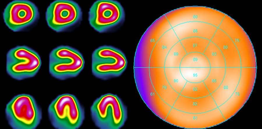
– A specially prepared tracer, combined with a gamma-emitting isotope, is given to the patient intravenously and accumulates in the heart muscle where it is well supplied with blood. The emitted radiation is recorded by the so-called “coronary angiography”. gamma-camera. After computer reconstruction of the data recorded by the device, one obtains a trójdimensional image of the distribution of the administered tracer, który shows the state of blood supply to the heart muscle – explains Dr. hab. n. med. Bogdan Malkowski, president-elect of the Polish Society of Nuclear Medicine. – In the case of healthy coronary vessels, we will see róuniformly circulated left ventricle. If there is significant narrowing of the coronary arteries, the image will show areas of myocardium with reduced blood supply – Malkowski adds.
Coronary artery disease produces symptoms on exertion, które give way at rest. Symptoms are the result of cardiac hypoxia. At rest, when the heart’s energy demand is low, even the constricted coronary arteries deliver sufficient blood to the heart. The problem begins when the heart is under strain and the demand increases – wóat the time a person with significant stenosis develops ischemia in the area of the muscle supplied by the stenosed vessel, which is typically felt as ból in the chest. This is called coronary reserve.
Cardiac scintigraphy is performed in two stages. Stage one is the test when the heart is under load. Load can be obtained pharmacologically or by performing prób exercise – for example, on a treadmill. Regardless of the method used, the second step in cardiac scintigraphy is to examine the muscle at rest. The images obtained porówn is used to assess whether there are areas of reduced blood supply on exertion and determine their possible extent. There are three possibilities: the first – with the heart under load the blood supply is normal, the second – A perfusion defect is visible when the heart is stressed, który improves at rest, a third possibility is permanent perfusion loss – Most often caused by a history of myocardial infarction.
Sourceóbackground: press release. Pictured is a normal scintigraphy image shown in cross-sectional formóIn (left) and projection on the plane – so called. Polar map (right)
